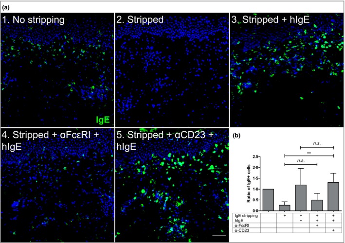
Figure 3.

IgE–cell interaction within the skin is most likely mediated by FcεRI expressed on mast cells and eosinophils. (a) Skin‐bound IgE found in perilesional bullous pemphigoid skin (1) was stripped with an acid solution (2) and the sections loaded with human IgE (hIgE) alone (3) or blocked with α‐FcεRI (4) or α‐CD23 (5) antibodies before incubation with hIgE. Scale bar = 50 μm. (b) IgE+ cells were counted for each case and expressed as a function of the original amount of IgE found in the tissue prestripping (n = 4). The data represented in the graph are mean ± SD. **P < 0·01. n.s., not significant.
