
Figure 4.
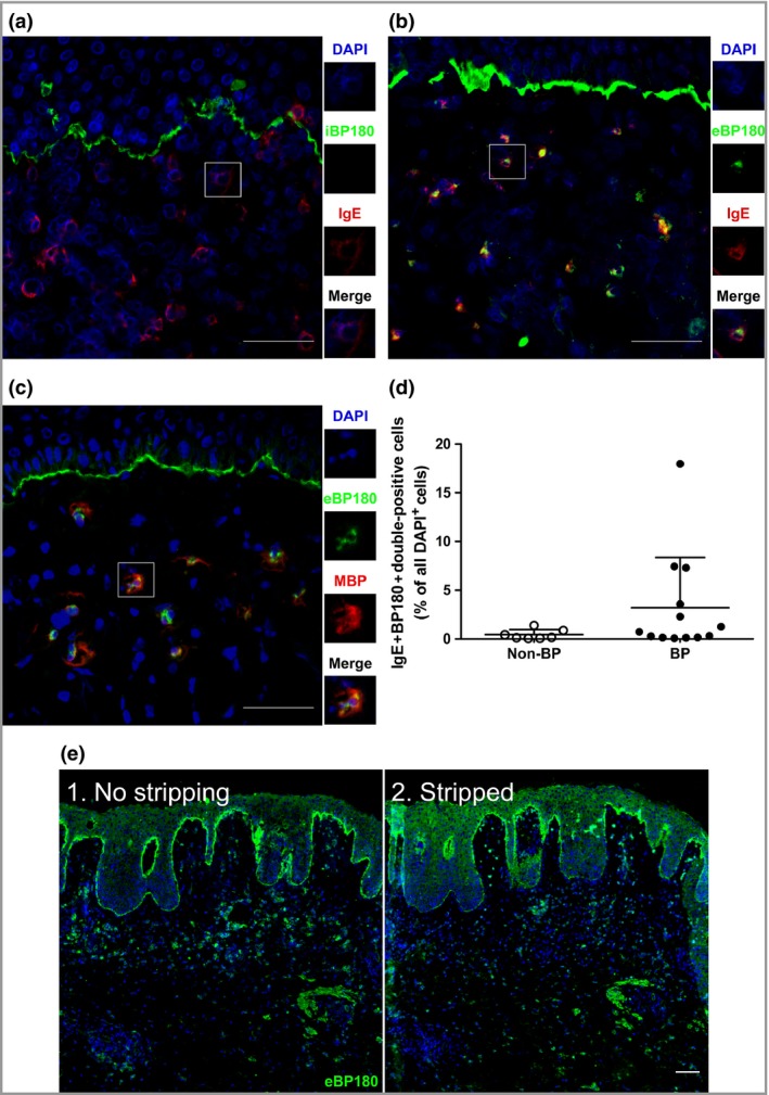
Shed fragments of BP180 are found in the dermis, colocalizing with IgE. Perilesional sections of skin from patients with bullous pemphigoid (BP) were stained for (a, b) IgE, (a) the intracellular moiety of BP180, (b, c) the extracellular moiety of BP180 and (c) major basic protein (MBP) (eosinophils). (d) The percentage of double‐positive (IgE+BP180+) cells, compared with all 4′,6‐diamidino‐2‐phenylindole (DAPI)+ dermal cells, in the skin of BP (n = 13) and non‐BP disease controls (n = 7), as stained by direct immunofluorescence (DIF). Non‐BP disease controls include linear IgA disease, vasculitis, epidermolysis bullosa acquisita, pemphigus and dermatitis herpetiformis. The data represented in the graph are mean ± SD. (e) DIF for extracellular BP180 moieties (eBP180) in a (1) nonstripped or (2) acid‐stripped BP section (n = 1). Scale bars = 50 μm.