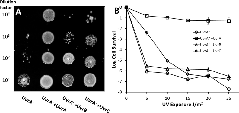
Figure 4.
Survival of UvrA− cells exposed to UV. (A) Spot plates of decreasing cell titers exposed to 5 J/m2 UV (254 nm) show improved survival with ectopic UvrB or UvrC. Lane 1 UvrA− cells, lane 2 UvrA− cells complemented with UvrA-eGFP, lane 3 UvrA− cells complemented with UvrB-eGFP, lane 4 UvrA− cells complemented with UvrC-eGFP. (B) Quantification of spot plates by colony counting. Survival of UvrA− cells complemented with eGFP-tagged NER proteins versus UV dose shows a significant improvement in survival at low UV doses (5–10 J/m2) for UvrC-complemented cells. Cell survival is shown in logarithm units and error bars indicate standard deviation.

