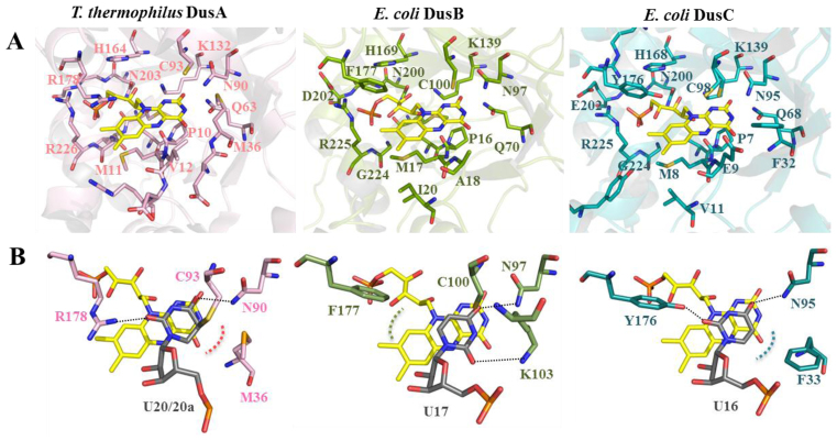
Figure 4.
Structure of Dus active site and uridine binding mode. (A) Structure of the FMN binding sites of Thermus thermophilus DusA (3B0P), Escherichia coli DusB (this study) and E. coli DusC (4BFA) in pink, green and blue, respectively. Residues involved in polar contacts with FMN (yellow stick) are shown as sticks. (B) Uridine binding mode in Dus active site as seen in the crystal structures of T. thermophilus DusA and E. coli DusC in complex with tRNA. In the case of E. coli DusB, the uridine was placed manually.

