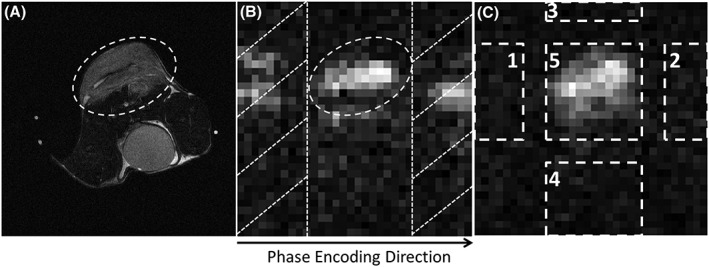
Figure 2.

Selection of ghost‐containing image areas, where the signal should be minimized using the proposed workflow. The tumour region was drawn from the proton image A, and the resulting region of interest (ROI) was then applied to the [1‐13C]lactate image B. Bands falling outside of the ROI in the phase‐encoding direction (bands with diagonal hatching in B) were considered as ghost‐containing background areas. The image in C, shows the selection of object 5 and background areas 1, 2, 3, 4 in one frame of the 13C images, which were used for the measurement of residual ghosting
