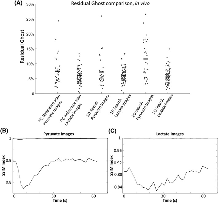
Figure 7.

A, Residual ghosting level in the lactate and pyruvate images acquired in vivo, where the images were phase corrected using a 13C reference scan, one‐dimensional (1D) search and two‐dimensional (2D) search. Both searches were performed on the [1‐13C]lactate image with the highest signal‐to‐noise ratio (SNR). The horizontal bars represent the mean value for that data cohort. The similarities (indicated with solid lines) between the images corrected with the 13C reference scan and the images corrected using a 1D search were measured for both the [1‐13C]pyruvate B, and [1‐13C]lactate C, images taken from a representative in vivo dataset [structural similarity (SSIM) indices ≈ 1]. The dotted lines show the similarities between the uncorrected images and the images corrected using reference scans
