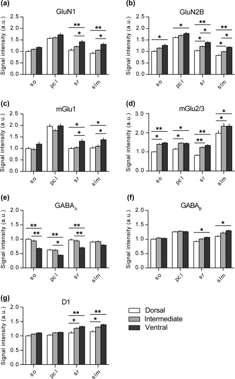
Figure 2.

Dorsal, intermediate and ventral parts of the CA1 region express different amounts of plasticity‐related receptors. Bar graphs illustrate the relative change in protein expression of the NMDAR subunits (GluN1 and GluN2B), group I and II mGlu receptors (mGlu1 and mGlu2/3), GABAergic receptors (GABAA and GABAB) and dopamine D1 receptors across the somato‐dendritic and longitudinal axes of the CA1 region. (a) GluN1 protein density levels were the highest in the ventral apical dendrites. (b) Similarly, the GluN2B levels were the highest in the ventral CA1 across its entire somato‐dendritic axis. Here, the protein expression in the apical dendrites showed gradual and significant increase from the dorsal towards the ventral pole. (c) Again, the mGlu1 protein density levels were the highest in the ventral apical dendrites of the CA1 region. (d) MGlu2/3 protein levels were the lowest in the dorsal CA1 and comparable in the intermediate and ventral domains. (e) GABAA protein expression was the lowest in the ventral CA1 in most layers and did not differ between the dorsal and intermediate parts. (f) GABAB levels were significantly higher in the ventral apical dendrites and were equally expressed in the cell soma and basal dendritic layers. (g) D1 protein levels were significantly higher in the ventro‐intermediate apical dendrites as opposed to their dorsal counterpart. Values expressed in arbitrary units. Error bars indicate SEM. *p < .05 or **p < .01. pcl, pyramidal cell layer; so, Stratum oriens; sr, Stratum radiatum; slm, Stratum lacunosum‐moleculare
