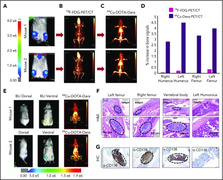
Figure 2.
64Cu-DOTA-Dara PET/CT shows higher resolution than does 18F-FDG-PET/CT. (A-C) Representative images of 2 NSG mice (mouse 1 and mouse 2), which were injected with 5 × 10⁶ MM.1S GFP+/Luc+ cells. After 2 weeks from the injection, the mice were imaged using BLI (A), and after 24 hours, the same mice were imaged by 18F-FDG-PET (B). The day after, 64Cu-DOTA-Dara (3.7 MBq, 10 µg) was administered to the same mice imaged with 18F-FDG-PET and BLI (C). (D) Graphical representation of the intensity of the signal of 64Cu-DOTA-Dara PET/CT in comparison with 18F-FDG-PET/CT, expressed as a percentage of the increase in percentage of the injected dose per gram (%ID/gr) of bone organs in comparison with the nonspecific signal of each radio nucleotide (%ID/gr) detected in the heart. A radiolabeled antibody identified MM cancer cells, which were undetectable by 18F-FDG-PET scan. (E) Mice with minimum BLI signals were IV-injected with 64Cu-DOTA-Dara (3.7 MBq, 10 µg) and then imaged by PET/CT, which identified MM cell dissemination in the bone (yellow boxes). (F-G) Representative images at different magnifications of tissue sections stained by hematoxylin and eosin stain (F) and CD138 immunohistochemical stain (G) showing MM cell accumulation (black dashed ovals) in the left and right femurs and in the vertebral body (lower lumbar), but not in the left humerus, which did not show PET/CT signals. H&E, hematoxylin and eosin; IHC, immunohistochemical.

