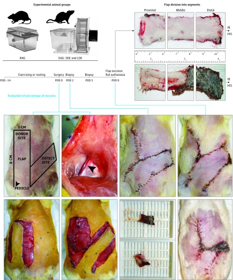
Figure 1. Principles of the Axial Fasciocutaneous Transposition Flap Animal Model and the Experimental Study Design.
Locations for biopsy excision are identified by gray squares at the bottom of each flap segment (seen on POD 2 image). Arrowheads indicate the pedicle and its vessel (superficial epigastric inferior artery and vein) bundle. Upper ruler shows centimeters, lower ruler shows inches. EAG indicates exercise animal group; HIS, histologic analysis; IB, immunoblotting; LDE, longer distance exercise; POD, postoperative day; RAG, resting animal group; and SDE, shorter distance exercise.

