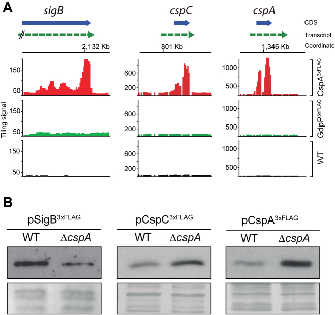
Figure 4.
Post-transcriptional regulation of selected CspA targets. (A) RIP-chip maps showing CspA-binding signals on the sigB, cspC and cspA loci. Normalized tiling signals of RIP-on-chip experiments for CspA3xFLAG, GdpP3xFLAG and WT are shown as red, green and black bars respectively. CDSs appear as a blue box arrow and mRNAs are represented as a dashed green arrow. (B) Expression of 3xFLAG-tagged SigB, CspC and CspA proteins in the WT and ΔcspA strains. Total protein extraction was performed at mid-exponential phase after growth at 37°C and 200 rpm. Samples were run into 12% polyacrylamide gels and transferred to nitrocellulose membranes. Western blots were developed using peroxidase conjugated anti-FLAG antibodies and a bioluminescence kit. Coomassie stained gel portions are shown as loading controls (LC).

