Figure 7.
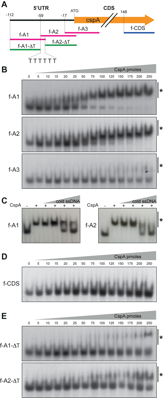
CspA binds to a T-rich motif in vitro. (A) Schematic representation of the ssDNA oligonucleotides designed to perform EMSAs with the recombinant CspA protein. (B) EMSA of f-A1, f-A2 and f-A3 ssDNA oligonucleotides (20 fmol of 32P-labeled synthetic oligo fragments) with increasing amounts of recombinant CspA protein. The pmoles per reaction used in each lane are indicated. (C) Gel shift competition assay of labeled f-A1 and f-A2 performed in the presence of increasing concentrations of unlabeled f-A1 and f-A2 and 25 and 50 pmol of recombinant CspA, respectively. (D) EMSA of the f-CDS ssDNA oligonucleotide (20 fmol of 32P-labeled synthetic oligo fragments) with increasing amounts of recombinant CspA protein. (E) EMSA of the f-A1-ΔT and f-A2-ΔT ssDNA oligonucleotides. These fragments lack the T-rich region from the 3′ and 5′ ends of f-A1 and f-A2, respectively. The CspA-oligonucleotide complexes are indicated with an asterisk.

