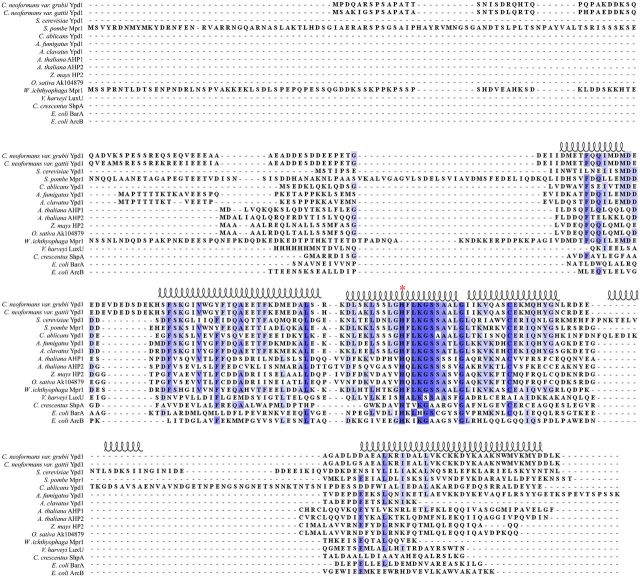
Figure 1.
Representative sequence alignment of HPt proteins. Amino acid sequences of HPt proteins were aligned using ClustalX 2.1 and visualized using Jalview 2.8.2. Highly conserved residues are shaded dark blue, while less conserved residues are shaded in lighter blue. The phospho-accepting histidine residue is denoted by a red asterisk (*). Secondary structure of the known crystal structure of Ypd1 from S. cerevisiae (PDB ID: 1QSP) is diagrammed above the alignment.

