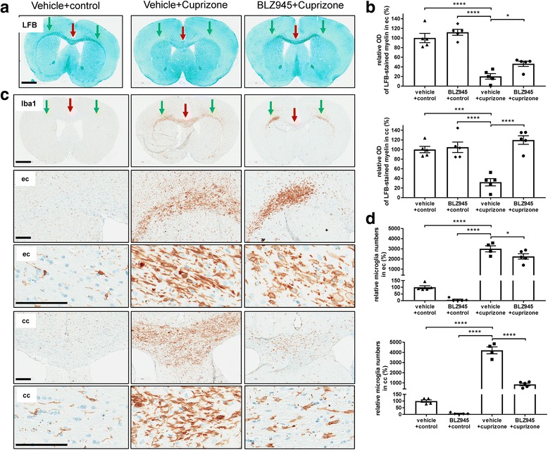
Fig. 6.

Prophylactic treatment with BLZ945 1 week before and during 5-week cuprizone intoxication inhibited demyelination and reduced microglia in the corpus callosum but enhanced axonal pathology and myelin debris in the external capsule. a Representative overview pictures from histological stainings of Luxol Fast Blue (LFB) for the different treatment groups at week5 (see Fig. 5a for the experimental setup and groups), red arrows: corpus callosum, green arrows: external capsule. b Corresponding analysis of the optical density (OD) of Luxol fast blue (LFB) in the cc and ec. c Representative overview and higher magnification pictures from immunohistological stainings detecting Iba1-positive microglia for the different treatment groups at week5 (see Fig. 5a for the experimental setup and groups), red arrows: corpus callosum, green arrows: external capsule. d Corresponding quantitative analysis of the immunohistochemistry for Iba1-positive microglia numbers in the corpus callosum and external capsule. Values were normalized to those of control vehicle mice. Group sizes: for all treatment groups n = 4–5. Data are shown as means±SEM. Scale bars: 200 μm for the higher magnification. Statistics: Turkey’s multiple comparison test one-way ANOVA (*: p < 0.05, ***: p < 0.001, ****: p < 0.0001), cpz: cuprizone, cc: corpus callosum, ec: external capsule, OD: optical density
