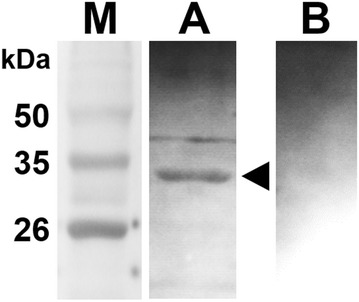
Figure 3.

Western blot analysis of purified LFliC. The protein was strongly recognized by mouse hyperimmune serum obtained from a mouse immunized with L. intracellularis. The arrow in lane A indicates a ~ 33 kDa band, which is the predicted size of LFliC fused with 6×His. Serum from an unimmunized mouse was used as a negative control (lane B). Lane M, size marker.
