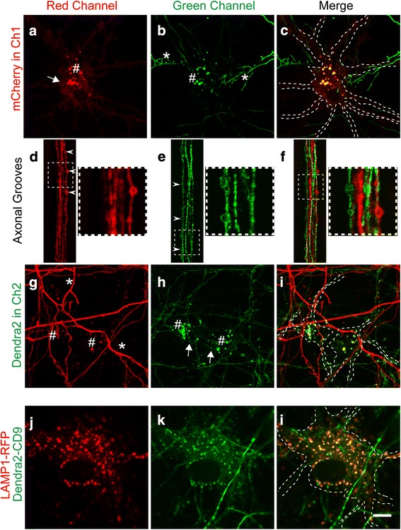
Fig. 2.

Transmission of exosomes between interconnected neurons. Culture performed according to Model 1, with neuron A being labeled with mCherry-CD9 (red) and neuron B labeled with Dendra2-CD9 (green). a-c Confocal images of mCherry-CD9-positive neurons in Ch1. The red channel (a) shows that CD9 is detected in the neuronal plasma membrane (a, white arrow) and is strongly present in endosomal somatic punctae (a, #). The green channel (b) reveals that Dendra2-positive axons (*) from Ch2 can reach mCherry-CD9-positive neurons in Ch1. Interestingly, red neurons only show green fluorescence in endosomal punctae when they are close to green axons (b, #). d-f Images of axonal grooves revealing that axons grow in both directions but never exchange fluorescence. White arrowheads indicate CD9-positive globular enlargements, which appear to be migrating endosomes based on single color fluorescence. The square (dashed lines) outlines a magnification of the axonal region (right square). f The magnification shows that the axonal endosomes are either green or red, but do not display both colors. This indicates that the fluorescence exchange found in the soma is post-synaptic. g-i Images of Ch2 containing Dendra2-CD9-expressing neurons. Similarly, Dendra2-expressing green endosomal punctae in Ch2 (h, #) show red fluorescence (g, #) when in proximity to red axons (g, *) projecting from Ch1. White arrows indicate the plasma membrane. j-l Colocalization of Dendra2-CD9-expressing endosomal punctae with the late endosome marker LAMP1 tagged with RFP (transduced with a baculovirus). Scale bar: 10 μm for all images
