Fig. 5.

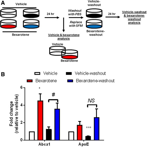
Abca1 remains significantly elevated while ApoE expression does not change after bexarotene removal in vitro. a Diagram representing the experimental procedure for analysis of bexarotene removal in primary astrocytes. Four groups of astrocytes were seeded: vehicle (white), bexarotene (red), vehicle-washout (black), and bexarotene-washout (blue). Vehicle (DMSO) or 10 nM bexarotene was applied to astrocytes in serum-free media (SFM) for 24 h after which vehicle and bexarotene samples were then used for protein analysis. Vehicle-washout and bexarotene-washout plates were then washed with PBS, serum-free media replaced, and incubated for an additional 24 h. Following this time point, vehicle-washout and bexarotene-washout samples were then used for protein analysis. b Quantification of each protein is represented as fold change relative to vehicle-treated cells. *p < 0.05 and ***p < 0.001, one sample t test with respect to vehicle-treated cells. Brackets indicate Student’s two-sample t test between indicated samples; #p < 0.05 and NS = not significant (exact p value = 0.111, Student’s t test with Welch’s correction for unequal variances). Data are representative of four separate experiments
