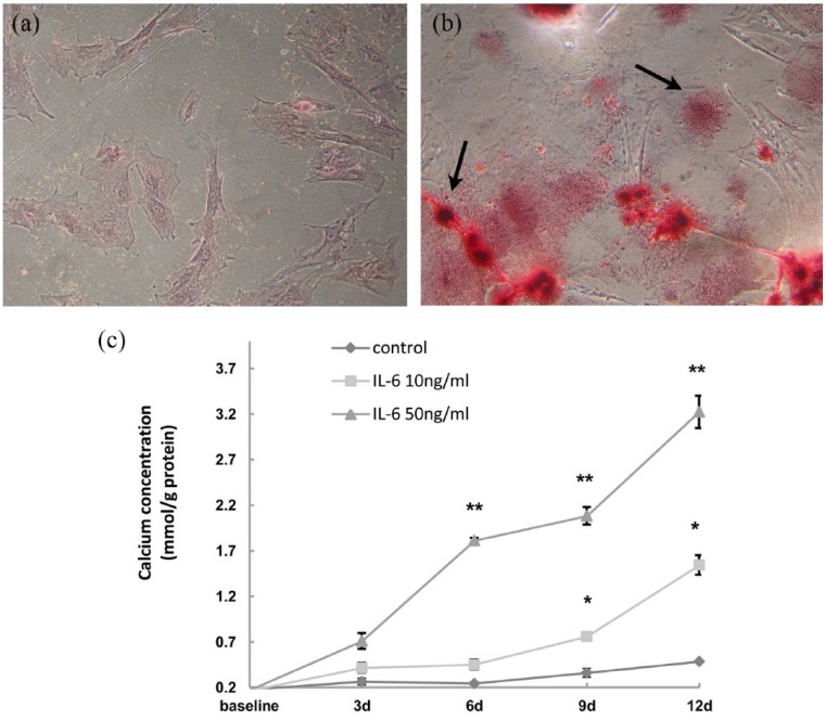
Figure 2.
IL-6 induced calcification of the HUASMCs. (a) The control group was treated without rhIL-6 for six days and showed a negative result of calcium stain by alizarin red S staining. (b) The experimental group was treated with rhIL-6 (50 ng/mL) for the same time period and the extracellular matrix was positively stained with orange. The two arrows indicated the sand-like and the nodular calcium stain in the extracellular matrix, respectively. Both panels were magnified 200 times. (c) Cells were treated with different concentrations of rhIL-6 for different durations as indicated. Calcium concentrations were elevated in a time- and dose-dependent manner. *P<0.05 vs. control of the same time point or vs. the sample of the earlier time point within the same group. **P<0.05 vs. control and sample of the other group at the same time point.

