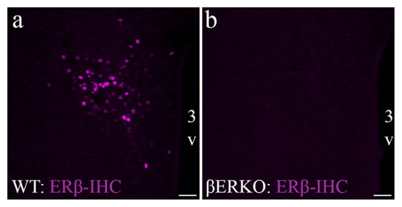
FIGURE 3.

ERβ Z8P antibody validation using the global ERβ knockout mouse model. Photomicrographs showing ERβ-ir in PVN of wildtype (WT; a) and global ERβ knockout (βERKO; b) following IHC using the Z8P antibody (magenta). Scale bars = 50 μm. 3V, Third ventricle [Color figure can be viewed at wileyonlinelibrary.com]
