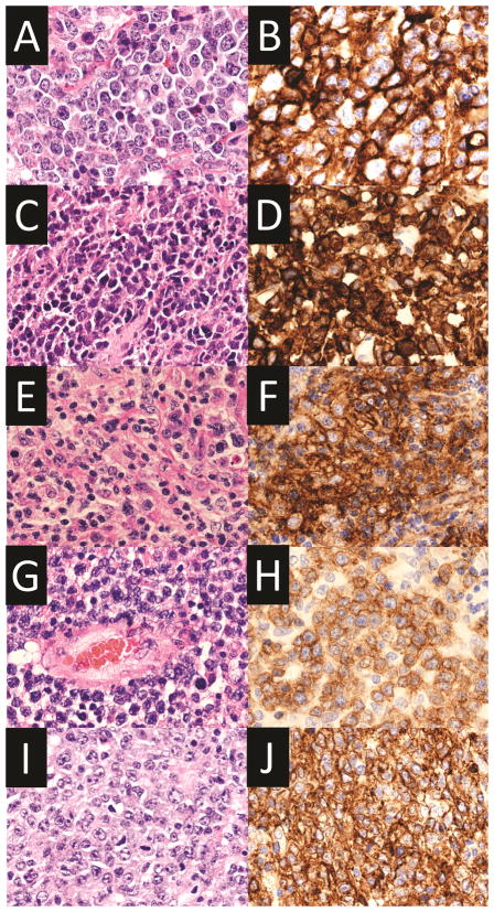
Figure 3.
Images of various lymphoid neoplasms with PD-L1 expression. A–B. Diffuse large B-cell lymphoma, not otherwise specified. Hematoxylin and eosin (A, x400) and PD-L1 stain (B, x400). C–D. Primary mediastinal large B-cell lymphoma. Hematoxylin and eosin (C, x400) and PD-L1 stain (D, x400). E–F. Epstein-Barr virus-positive diffuse large B-cell lymphoma, not otherwise specified. Hematoxylin and eosin (E, x400) and PD-L1 stain (F, x400). G–H. Primary central nervous system lymphoma. Hematoxylin and eosin (G, x400) and PD-L1 stain (H, x400). I–J. Primary testicular lymphoma. Hematoxylin and eosin (I, x400) and PD-L1 stain (J, x400). All PD-L1 stains were performed using SP142 clone (Spring Bioscience, Pleasanton, CA, USA).

