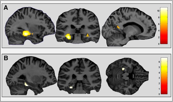
Figure 3.

Brain regions responsive to sentence type. A, PHC, extending into hippocampus (left and middle) and a small cluster in RSC (right) showed greater activity for imageable sentences (categories 1–4) than non-imageable sentences (categories 5 and 6). B, Left PHC showed greater activity for imageable sentences describing a landmark (categories 1 and 2) than those describing an action (categories 3 and 4). Activations are shown on views from a single representative participant's structural MRI brain scan, displayed at a whole-brain FWE-corrected threshold of p < 0.05. The color bar indicates the Z-scores associated with each voxel.
