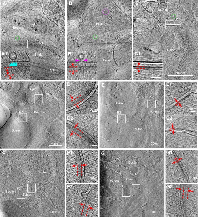
Figure 2.
Synapses of various sizes, shapes, and ultrastructural details imaged with cryo-ET. A–C, Three tomographic slices showing structures of different synapses. In the synapses, structures, such as SVs and dense core vesicle in presynaptic boutons (Bouton), microtubules (MT) in boutons and dendritic shaft (Shaft), mitochondria (Mit) in presynaptic bouton and postsynaptic spine (Spine), are clearly visible. A1–C1, Zoomed-in views of corresponding boxed areas from A–C showing thick (A1, B1, dashed parallel lines) and thin (C1, dashed parallel lines) PSDs, as well as SVs attached (A1, cyan arrowhead) or fused (B1, pink arrowheads) to the presynaptic membrane. D, E, Two synapses sharing the same presynaptic axon (determined by following through their tomograms in 3D), both with thick PSDs (D1 and D2) or both with thin PSDs (E1 and E2), respectively. F, G, Two synapses sharing the same postsynaptic spine, both with thick PSDs (F1 and F2), or one with thin PSD (G1) and the other with thick PSD (G2).

