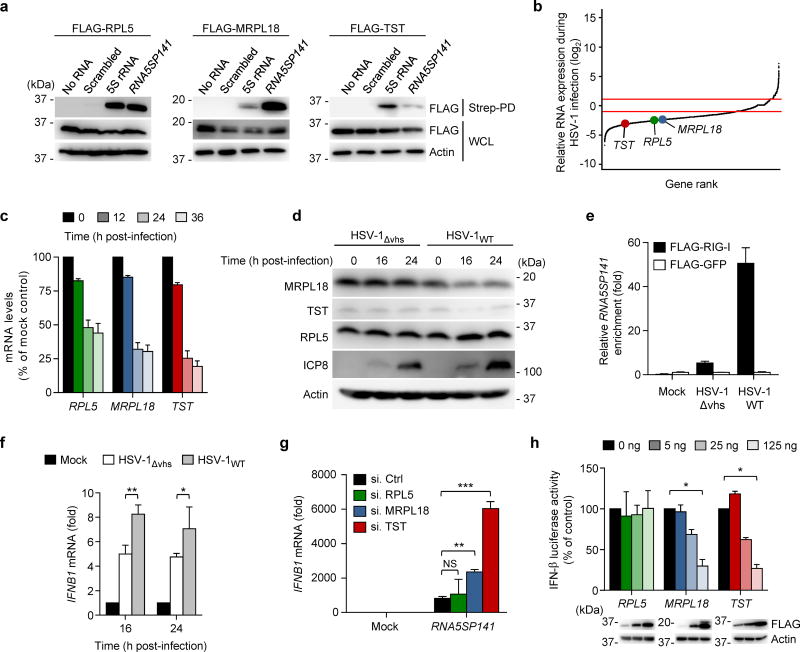
Figure 4.
Downregulation of TST and MRPL18 by HSV-1 allows RIG-I activation. (a) Binding of biotinylated in vitro-transcribed RNA5SP141 to FLAG-tagged RPL5, MRPL18, and TST in transiently transfected HEK 293T cells, assessed by streptavidin pulldown (Strep-PD) and IB with anti-FLAG. WCLs were probed by IB with anti-FLAG and anti-actin. Biotinylated 5S rRNA and a scrambled random RNA (Scrambled) served as positive and negative controls, respectively. (b) Relative mRNA expression of RPL5, MRPL18, and TST in HEK 293T cells infected with HSV-1 (MOI 1) for 16 h as compared to uninfected cells, determined by RNAseq. Red boundaries represent ±2-fold change in gene expression. (c) qRT-PCR analysis of RPL5, MRPL18, and TST mRNA in HEK 293T cells infected with HSV-1 (MOI 1) for the indicated times. (d) IB analysis of endogenous RPL5, MRPL18, and TST proteins in the WCLs of HEK 293T cells infected with HSV-1WT (revertant) or HSV-1Δvhs (both MOI 10) for the indicated times. IB analysis of HSV-1 ICP8 and cellular Actin served as infection and loading controls, respectively. (e) HEK 293T cells were transfected with FLAG-RIG-I or FLAG-GFP. 24 h later, cells were infected with HSV-1WT (revertant) or HSV-1Δvhs (both MOI 10) for 16 h, or left uninfected (Mock). RNA bound to FLAG-RIG-I or FLAG-GFP was precipitated from cell lysates using anti-FLAG PD as described in Figure 1a, followed by qRT-PCR analysis to assess bound RNA5SP141 transcripts. (f) qRT-PCR analysis of IFNB1 mRNA in NHLF cells infected with HSV-1WT (revertant) or HSV-1Δvhs (both MOI 1) for 16 h and 24 h, or left uninfected (Mock). (g) qRT-PCR analysis of IFNB1 transcripts in HEK 293T cells transfected with the indicated siRNAs for 30 h and then transfected with either no RNA (Mock) or 1 pmol of in vitro–transcribed RNA5SP141 for 16 h. (h) IFN-β luciferase reporter activity in HEK 293T cells transfected for 30 h with the indicated amounts of plasmids expressing FLAG-tagged RPL5, MRPL18, or TST and subsequently transfected with 1 pmol of RNA5SP141 for 16 h to stimulate RIG-I signaling. Expression of FLAG-tagged proteins was confirmed in the WCL by IB with anti-FLAG. Data are representative of two (a, c-f), one (b) or three (g, h) independent experiments (mean and s.d. of n = 3 technical replicates in e, n = 2 biological replicates in c, f, and h, or n = 3 biological replicates in g). *P < 0.05, **P < 0.01, ***P < 0.001 (unpaired t-test). NS, statistically not significant.

