
Figure 5.
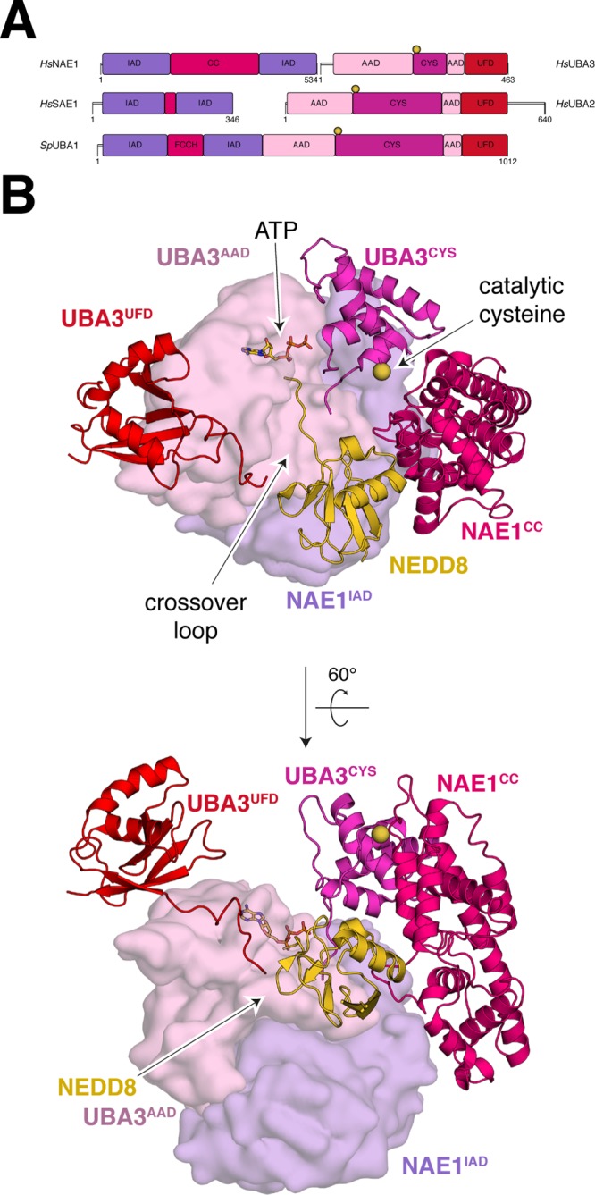
Domain organization of canonical E1s. (A) Primary structure of canonical E1s for NEDD8, SUMO, and ubiquitin. (B) Structure of human NEDD8∼E1NAE1/UBA3/E2UBC12/ATP (PDB 2NVU). Adenylation domains are shown in a Gaussian surface representation, while other domains are in cartoon representation. The sulfur atom of the active site cysteine is in sphere representation colored yellow. Positions for ATP, the crossover loop, and the active site cysteine are highlighted by arrows.