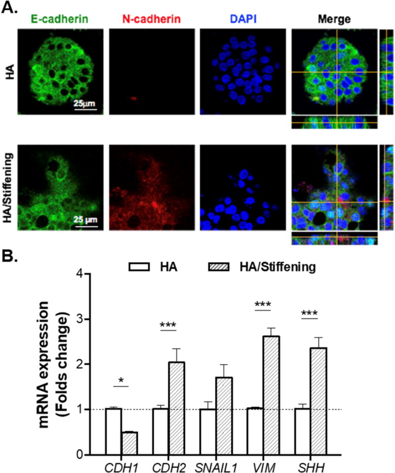
Figure 9. Evaluation of selected epithelial and mesenchymal markers in COLO-357 cells grown in HA-containing and soft (i.e., Gel/HA) or in HA-containing and TYR-stiffened gels (i.e., GelHPA/HA).

(A) Immunofluorescence staining of E-cadherin and N-cadherin. Cells were counterstained with DAPI. (B) mRNA expression levels of CDH1 (E-cadherin), CDH2 (N-cadherin), SNAIL1, VIM (vimentin), and SHH (sonic hedgehog). All assays were conducted with samples collected at day 14. (Housekeeping gene: GAPDH. N=3, Mean ± SEM. *p<0.05, ***p<0.001).
