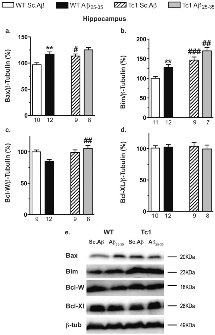
Figure 8.
Analyses of apoptotic pathway in the hippocampus of Tc1 mice injected with amyloid-β [25-35] (Aβ25-35) peptide. Wildtype (WT) or Tc1 mice were administered intracerebroventricular (i.c.v.) injections with scrambled peptide (Sc.Aβ) or Aβ25-35 peptide (9 nmol) and sacrificed 10 days after injection. The levels of pro-apoptotic proteins, Bax (a) and Bim (b), and anti-apoptotic proteins, Bcl-W (c) and Bcl-XL (d), were assessed by Western blot in the hippocampal protein lysates. Typical blots are shown in (e). The number of animals per group is indicated below the columns. Two-way analysis of variance (ANOVA): F(1,35)=8.37, p<0.01 for the genotype, F(1,35)=14.1, p<0.001 for the treatment, F(1,35)=1.03, p>0.05 for the interaction in (a); F(1,36)=34.8, p<0.0001 for the genotype, F(1,36)=10.1, p<0.01 for the treatment, F(1,36)=0.559, p>0.05 for the interaction in (b); F(1,33)=6.28, p<0.05 for the genotype, F(1,33)=1.22, p>0.05 for the treatment, F(1,37)=7.71, p<0.01 for the interaction in (c); F(1,30)=0.463, p>0.05 for the genotype, F(1,30)=1.47, p>0.05 for the treatment, F(1,30)=1.48, p>0.05 for the interaction in (d); *p<0.05; **p<0.01 vs same genotype Sc.Aβ-treated mice; #p<0.05; ##p 0.01; ###p<0.001 vs same treatment WT mice; Bonferroni’s test.

