
Figure 3.
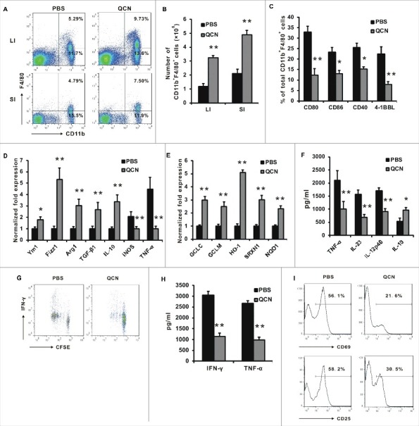
QCN induces the functional alteration of enteric macrophages in experimental colitis. (A–B) The frequency (A) and cell number (B) of CD11b+ macrophages isolated from the large intestine (LI) and small intestine (SI) in naïve CD4+ T-cell-mediated colitis. (C) Phenotypic characterization of macrophages from the colon in CD4+ T-cell-mediated colitis. (D–E) Real-time PCR for the expression of genes in CD11b+ F4/80+ sorted from pooled colonic LPL in CD4+ T-cell-mediated colitis. (F) Cytokine levels in the LPS-stimulated colonic macrophages that were collected at day 45 following T-cell transfer. (G–H) CFSE-labeled OT-II T cells were co-cultured with PBS or QCN-pretreated lamina propria macrophages and OVA peptide (5 µg/mL). Proliferation of CFSE-labeled CD4+ T cells (G) and the level of IFN-γ and TNF-α (H) in the supernatant of co-cultured cells were examined. T-cell activation molecules (I) were analyzed by FACS after 4 days in culture. Data (B–D, H) are presented as the mean ± SEM. *P<0.05, **P<0.01 (Student's t-test).