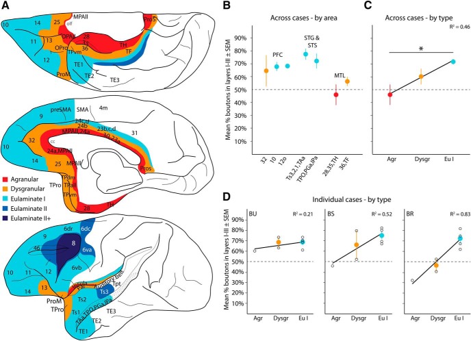
Figure 10.
Laminar patterns of A25 pathways vary by cortical type: axon terminations. A, Map of cortical areas connected with A25 are color-coded by cortical type: agranular (red) to eulaminate II+ (darkest blue). Areas outside the PFC with no A25 connections were left blank. B, Average percentage boutons across cases from A25 (±SEM) found in superficial layers (Cases BU, BS, BR; Case BP had only a retrograde tracer). C, Cortical areas from B were pooled across cases and regressed by cortical type. This analysis showed that the proportion of labeled boutons in the superficial layers increased as cortical structure (type) became more elaborate. D, Individual cases with average percentage of labeled boutons in superficial layers (±SEM) and linear fit from regression show a consistent upward trend. Black circles represent individual cortical areas and convey variability. olf, Olfactory cortex and nuclei. *significant difference.

