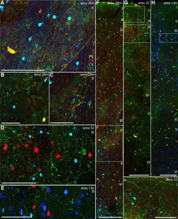
Figure 11.
Examples of A25 axon terminations within distinct inhibitory microenvironments. A–C, High magnification of layer V in medial entorhinal area 28 shows A25 terminations (green) comingling with PV neurons and their processes (yellow) and with deeply situated CR neurons (blue). A, Inset from F. D, High magnification of layer III in area 32 shows A25 terminations comingling with CB (red) and CR (blue) neurons (inset from G). E, High magnification of layer III in area 12o shows A25 terminations comingling with CB (red) and CR (blue) neurons (inset from H). F, Low magnification of column through the entire depth of medial entorhinal area 28 shows A25 terminations (green) seen mostly in layer V where PV (yellow) and CR (blue) inhibitory neurons are found (Case BU). G, Low-magnification column through the cortical depth of area 32 shows A25 terminations in the superficial layers, where CB (red) and CR (blue) neurons are prominent (Case BR). H, Low magnification of column through the cortical depth of orbital area 12 shows terminations from A25 mostly in the upper layers, where CB (red) and CR (blue) neurons are found (Case BU). I, High magnification of layer I in area 32 shows A25 terminations coursing through layer I, which contains the apical dendrites from pyramidal neurons below (inset from G). Layer I contains almost no cell bodies, with the exception of CR neurons. Empty arrowheads indicate A25 terminations (green) or color-coded processes of inhibitory neurons. Filled green arrowheads indicate labeled projection neurons directed to A25. Scale bars: A, D, E, I, 100 μm; B, C, 50 μm; F–H, 200 μm. ld, Lamina dissecans.

