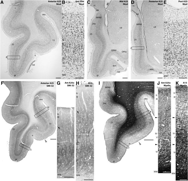
Figure 2.
Architecture of A25. A, Nissl-stained coronal section through anterior A25 (Case BB). B, Higher-magnification inset of anterior medial A25. C, Nissl-stained coronal section through mid-level A25. D, Nissl-stained coronal section through posterior A25. E, Higher-magnification inset of posterior medial A25. F, Coronal section stained for SMI-32 through anterior A25 and area 13 (Case BB). Gray arrowhead indicates the transition from A25 to area 13. G, Higher-magnification inset of anterior orbital A25. H, Higher-magnification inset of area 13. Black arrowheads indicate the increase in SMI-32-labeled neurons and processes in layer III of area 13. I, Coronal section stained for myelin through anterior A25 and area 13 (Case AN). Gray arrowhead indicates the transition from A25 to area 13. J, Higher-magnification inset of anterior orbital A25. K, Higher-magnification inset of area 13. Black arrowheads indicate the increase in myelin labeling in area 13 layers III–VI. Scale bars: regional photomicrographs, 1 mm; column insets, 250 μm. All, Allocortex; aon, anterior olfactory nucleus; cc, corpus callosum; cd, caudate; gr, gyrus rectus; lot, lateral olfactory tract; mo, medial orbital sulcus; OPAll, orbital periallocortex (agranular); OPro, orbital proisocortex (dysgranular); o, olfactory sulcus; WM, white matter; 25m, medial A25; 25o, orbital A25.

