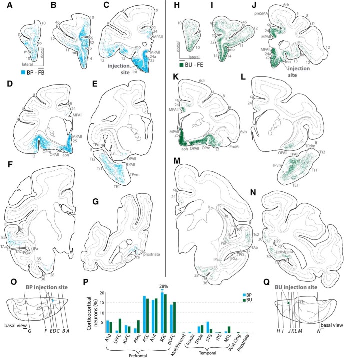
Figure 4.
Distribution of cortical labeled neurons directed to orbital A25. A–N, Coronal sections from rostral (A, H) through caudal (G, N) levels show distribution of retrogradely labeled neurons across cortical areas in 2 cases (A–G: Case BP; H–N: Case BU). Dotted lines on coronal sections indicate boundary between superficial and deep layers as delimited by the bottom of layer III. Most labeled projection neurons were found along the orbital and medial surfaces (B–D, I–K). O, Q, Injection sites on the basal surface of the brain and the level of the coronal sections depicted above. Scale bars, 2 mm. P, Proportion of labeled neurons found by cortical region (grouping of areas is shown below). There is striking similarity of the 2 A25 cases by injection site and distribution of labeled neurons. Grouped areas are as follows: LPFC, areas 46, 9l, 8, 12l; aOFC, 11, 12o; ACC, 32, perigenual 24a and MPAll; SGC, 25, subgenual 24a and MPAll; pOFC, 13, OPro, OPAll; Mot/Premot, 6, preSMA, SMA, 4; Insula, Ia, Id; TPole, TPro, TPAll, TPdm, TPvm; STG, PaI, PaAr, PaAlt, Ts3, Ts2, Ts1, Tpt, MST, TAa, TPO, PGa, IPa, FST; ITG, TEa, TEm, TE1, TE2, TE3, TEO; MTL, areas 28, 35, 36, TH, TF; Post Cing, 23, 30; Prostriata, agranular and dysgranular prostriata. a, Arcuate sulcus; aon, anterior olfactory nucleus; ca, calcarine sulcus; cg, cingulate sulcus; FPole, frontal pole; Ia, agranular insula; Id, dysgranular insula; ITG, inferior temporal gyrus; lf, lateral fissure; lo, lateral orbital sulcus; lot, lateral olfactory tract; mo, medial orbital sulcus; OPAll, orbital periallocortex; OPro, orbital proisocortex; p, principal sulcus; ProM, motor proisocortex; rh, rhinal sulcus; ro, rostral sulcus; st, superior temporal sulcus; TPAll, temporal periallocortex; TPdm, temporal proisocortex dorsomedial; TPvm, temporal proisocortex ventromedial.

