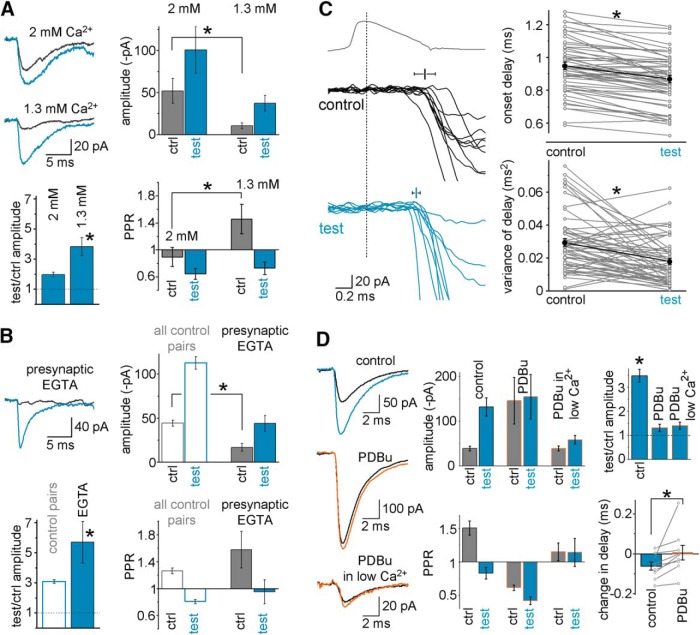
Figure 5.
Investigations into mechanisms of postburst plasticity in FF-INs. A, Persistence of the postburst potentiation in lower (1.3 mm; allegedly physiologically more relevant) extracellular Ca2+ levels. Example traces are shown from a CA3 GC-IvyC pair in which the effects of single presynaptic bursts were first tested in the presence of standard 2 mm Ca2+ concentrations and then in 1.3 mm Ca2+ (preburst control responses: black; test responses: blue, 3.6 s after burst). Bar graphs represent average data from all pairs (n = 8); the lower Ca2+ concentration decreased the response amplitudes and increased the PPRs, but the potentiation persisted. B, Example traces from a CA3 GC-AAC pair in which the effects of single presynaptic bursts were tested while the presynaptic CA3 GC was recorded with 1 mm intracellular EGTA to chelate Ca2+. Bar graphs represent average data from all presynaptic EGTA pairs (n = 8). Presynaptic EGTA strongly decreased the control responses, but the burst-induced potentiation persisted. C, Example traces illustrate the accelerated and more precise release before and after single presynaptic bursts from the same pair; average presynaptic APs and individual EPSCs are shown; mean and variance of the delay are also indicated, with error bars; failures were excluded for clarity. Right panels, Plots of the mean and variance values of the synaptic onset delay in each MF-FF-IN pair before and after single bursts (connected gray symbols) and their average (black). D, The DAG analog phorbol ester PDBu (1 μm), which promotes vesicle priming, increased MF-EPSC amplitude before bursts (note the different scale bars) and prevented burst-induced potentiation in a representative pair. Subsequent reduction of the release probability following the application of decreased extracellular Ca2+ (1 mm) in the PDBu-containing perfusing solution significantly reduced the average amplitudes, but burst-induced amplification remained negligible in the same pair. Summary bar graphs show that bursts were no longer effective in eliciting potentiation in the same pairs in the presence of PDBu regardless of the release probability; furthermore, PDBu also prevented the burst-induced decrease in the delay of the responses (bottom right). Symbols represent changes in synaptic delays in control conditions and in the presence of PDBu. Bars represent average data. * marks significant difference.

