Figure 3.

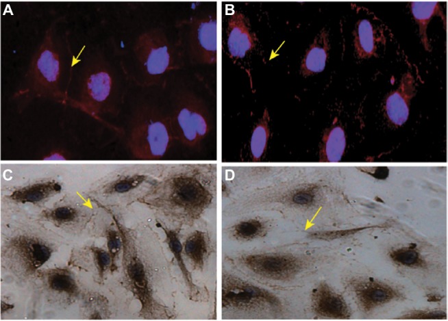
ZO-1 expression in HBMVECs.
Notes: (A) ZO-1 expression in HBMVECs before exposure to leukemia cells, as determined by immunofluorescence microscopy. (B) ZO-1 expression in HBMVECs after exposure to leukemia cells, as determined by immunofluorescence microscopy. (C) ZO-1 expression in HBMVECs before exposure to leukemia cells, as determined by immunohistochemistry. (D) ZO-1 expression in HBMVECs after exposure to leukemia cells, as determined by immunohistochemistry. ZO-1 immunoreactivity (red staining) was restricted to junctional areas and some punctate staining in the cytoplasm (arrows). Exposure to leukemia cells resulted in a weaker junctional and cytoplasmic pattern of ZO-1 immunoreactivity. DAPI (blue staining) was used to visualize the nuclei. Magnification: 1000×.
Abbreviations: HBMVECs, human brain microvascular endothelial cells; ZO-1, Zonula occludens-1; DAPI, 4′,6-diamidino-2-phenylindole.
