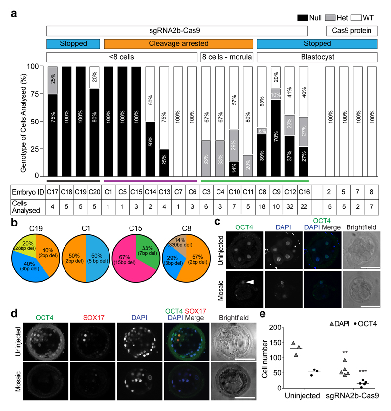
Figure 3. Genotypic characterization of OCT4-targeted human embryos.
a, Proportion of POU5F1-null, heterozygous or wild-type cells in each human embryo. The number of separate individual cells analysed is indicated. Embryos 2, 5, 7 and 8 were microinjected with Cas9 protein as a control. All other embryos were microinjected with the sgRNA2b–Cas9 ribonucleoprotein complex. The development of some embryos was stopped and they were removed from culture for analysis, while others were analysed following cleavage arrest. b, The types and relative proportions of indel mutations observed compared to all observable indel mutations within each human embryo. c, Immunofluorescence analysis for OCT4 (green) and DAPI nuclear staining (blue) in an uninjected control cleavage stage human embryo or an embryo that developed following sgRNA2b–Cas9 microinjection (n = 5). Confocal z-section. Arrowhead, OCT4-expressing cell. Scale bar, 100 μm. d, Immunofluorescence analysis for OCT4 (green), SOX17 (red) and DAPI nuclear staining (blue) in an uninjected control human blastocyst (n = 3) or a blastocyst that developed following sgRNA2b–Cas9 microinjection (n = 3). Confocal z-section. Scale bar, 100 μm. e, Quantification of the number of DAPI- or OCT4-positive nuclei in uninjected control human blastocysts (n = 3) compared to blastocysts that developed following sgRNA2b–Cas9 microinjection (n = 5). One-tailed t-test. **P < 0.01; ***P < 0.001.

