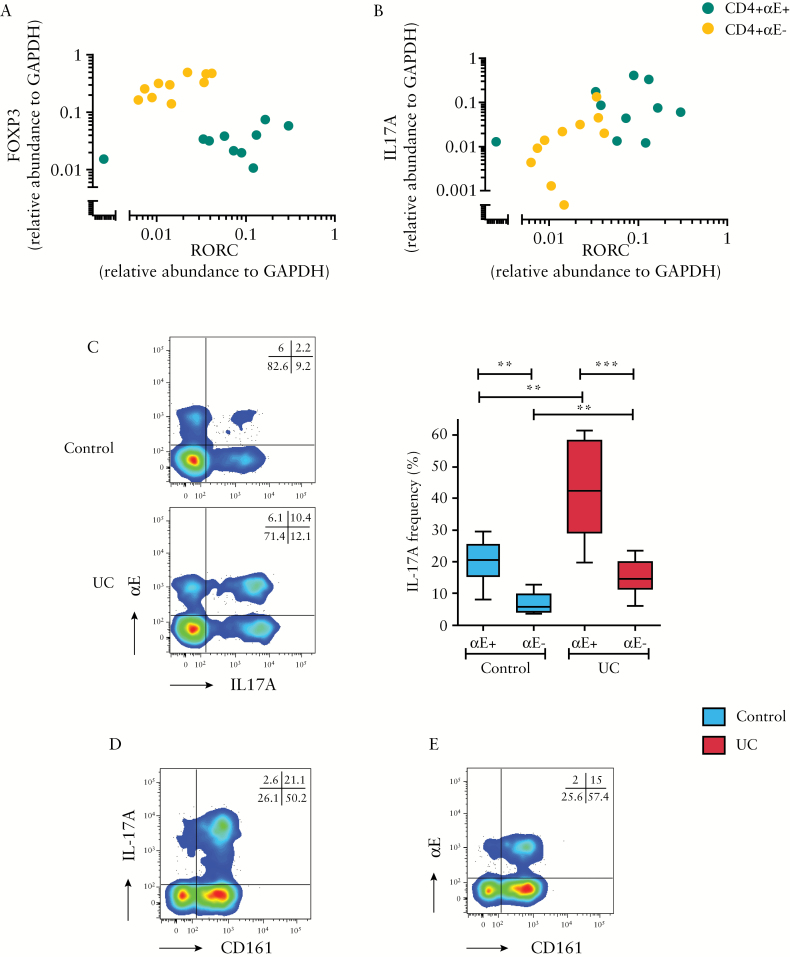
Figure 3.
αE integrin expression by colonic CD4+ T lymphocytes is associated with Th17 differentiation. FACS-sorted, unstimulated CD4+αE+ and CD4+αE− T lymphocytes isolated from active UC colonic biopsies were analysed for gene expression of [A] FOXP3 vs RORC and [B] IL17A vs RORC [n = 10 patients]. [C] In a separate study cohort, IL-17A protein expression in CD4+αE+ and CD4+αE− T cells was evaluated by FACS in colonic ex-vivo stimulated T lymphocytes from control subjects [n = 6] and patients with active UC [n = 8]. Representative two-colour FACS plots from control and UC patients are shown alongside summary Tukey box plots. Representative flow cytometry showing CD4+ T cell CD161 co-expression with [D] intracellular IL-17A, and [E] surface αE staining on a patient with active UC.

