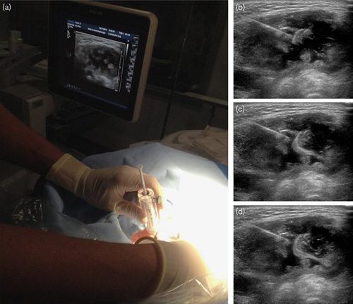
Fig. 1.

(a) Setup of the ultrasound-guided injection with the syringe with holmium-166 microspheres shielded with acrylic glass. (b–d) Ultrasound images of an injection in a large necrotic fluid-filled lymph node metastasis, clearly visible flow of microspheres inside the tumor.
