Fig. 3.

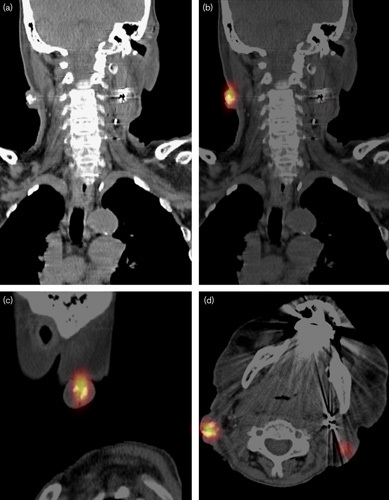
Patient 3, (a) clearly visible accumulations of holmium-166 microspheres as white dots on computed tomography. (b–d) Single-photon emission computed tomography reconstructions in coronal, sagittal, and axial directions.

Patient 3, (a) clearly visible accumulations of holmium-166 microspheres as white dots on computed tomography. (b–d) Single-photon emission computed tomography reconstructions in coronal, sagittal, and axial directions.