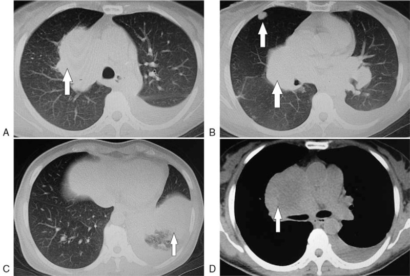
Figure 1.

Metastatic nidi on the chest computed tomography images taken in March 2016. A: Right hilar metastatic nidi, B: Right hilar and intrapulmonary metastatic nidi, C: Left lower pulmonary obstructive pneumonia, and atelectasis, D: Right hilar and mediastinal lymph node metastases.
