
Figure 2.
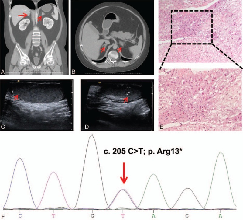
Results of imaging, histopathology, and DNA sequencing. Adrenal computed tomography scan showed bilateral adrenal nodular hyperplasia (A and B). Ultrasonography demonstrated multiple microcalcifications of the bilateral testes (C and D). (E) Hematoxylin and eosin staining of the adrenal tissue revealed multiple pigmented nodules. (F) Sequencing of DNA extracted from peripheral leukocytes identified a heterozygous C>T substitution in PRKAR1A exon 3 (indicated by the red arrow).