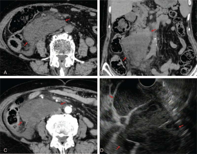
Figure 1.

Idiopathic retroperitoneal abscess on CT and EUS. A and B, A 9.1 cm × 4.2 cm × 11.7 cm large mass in the retroperitoneal cavity below the right kidney and horizontal portion of the duodenum was revealed on unenhanced CT (surrounded by red arrows). C, A hypoenhanced mass was revealed on contrast-enhanced CT (in between the red arrows). D, EUS showed the peripheral rim of the abscess, solid necrotic structure, and partition wall inside the lesion (in between the red arrows). CT= computed tomography, EUS= endoscopic ultrasound.
