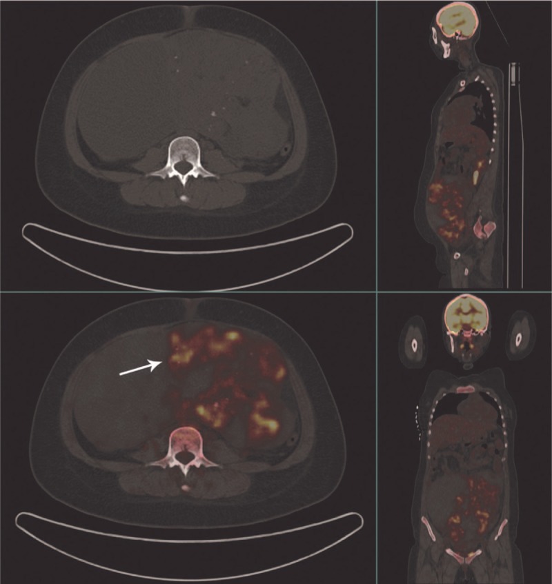
Figure 1.

Before surgery, CT images demonstrated a large mass with a maximum diameter of 25 cm with solid, cystic, fat, and calcified components. 18F-FDG PET/CT showed pathological FDG uptake in solid components of the abdominopelvic mass. Intensely increased FDG uptake was also seen in the retroperitoneal lymph nodes of the bilateral pelvic wall and bilateral iliac fossa. CT = computed tomography, 18F-FDG PET/CT = fluorine-18 fluorodeoxyglucose positron emission tomography/computed tomography.
