Figure 1.

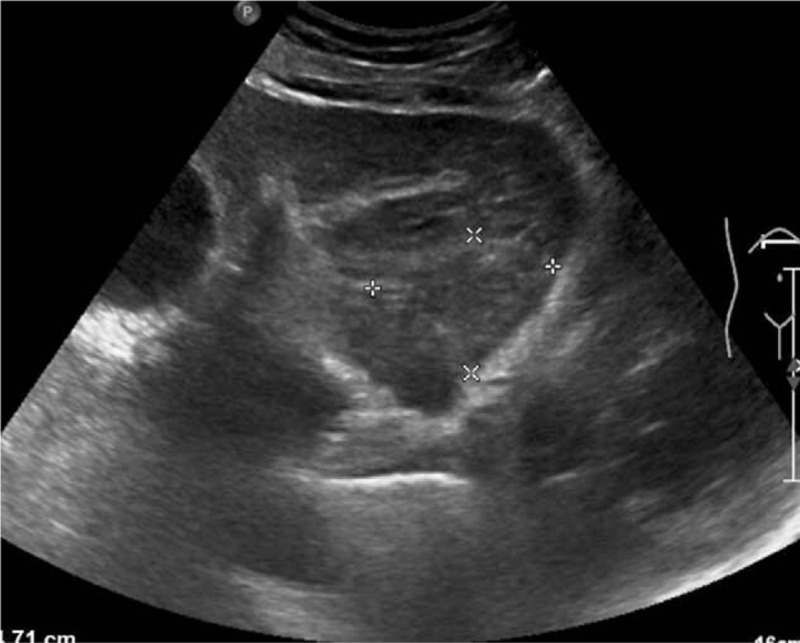
Abdominal ultrasonography. A well-defined, heterogeneously hypo-echoic mass (4.7×3.6 cm in size) was found in the left lateral liver. The boundary and vascular network were not clearly defined.

Abdominal ultrasonography. A well-defined, heterogeneously hypo-echoic mass (4.7×3.6 cm in size) was found in the left lateral liver. The boundary and vascular network were not clearly defined.