Figure 1.

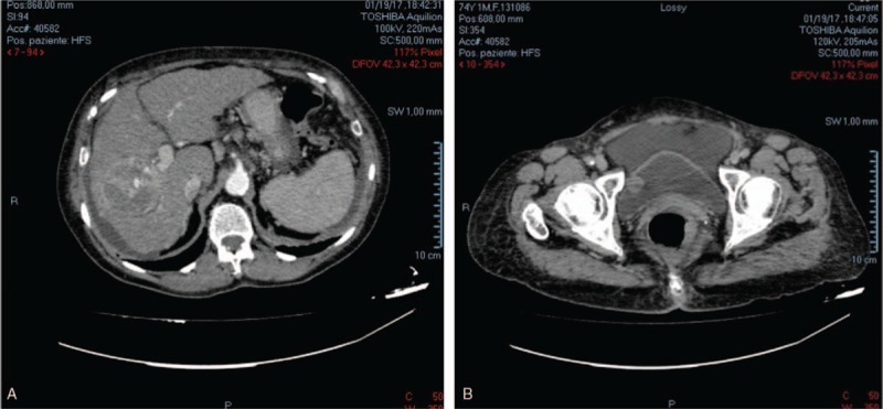
Abdominal computed tomography (CT) scanning showing: (A) a 68 mm liver nodule with nonhomogeneous contrast enhancement and a central necrosis area suspected for carcinoma; (B) an 18 mm bladder neoplasm of right bladder wall.

Abdominal computed tomography (CT) scanning showing: (A) a 68 mm liver nodule with nonhomogeneous contrast enhancement and a central necrosis area suspected for carcinoma; (B) an 18 mm bladder neoplasm of right bladder wall.