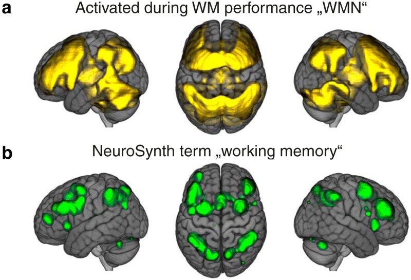
Figure 1.

WMNs. A, Brain regions that were more strongly activated during the 2-back condition in comparison to the 0-back condition in our sample (2-back – 0-back contrast one-sample t tests FDR corrected, α = 0.05). B, Meta-analytic results for the term working memory retrieved from NeuroSynth (reverse inference, FDR corrected, α = 0.01). The brain images are displayed within the MNI152 template and according to neurologic convention.
