. 2018 Jan 29;7:e32319. doi: 10.7554/eLife.32319

Figure 4. olMG cells divide in the INL with an apico-basal spindle orientation.

(A–A’’) Cryosection of an injured hatchling medaka retina of the transgenic line rx2::H2B-eGFP. PH3 stainings (magenta) on hatchling medaka retinae 3 days post PRC injury show mitotic olMG cells present in the INL (arrowhead), co-localizing with the rx2 nuclear reporter expression (green) (n = 4 fish, data obtained from three independent experiments). (B–B’’’) In vivo imaging of hatchling rx2::H2B-eGFP medaka retinae which were injured in the ONL and imaged starting at 44 hpi. OlMG nuclei which start to condense their chromatin can be detected in the INL (arrowheads). The divisions occur in an apico-basal manner (n = 6 fish, data obtained from six independent experiments, 5 out of 6 imaged divisions were apico-basal). Scale bars are 10 μm.

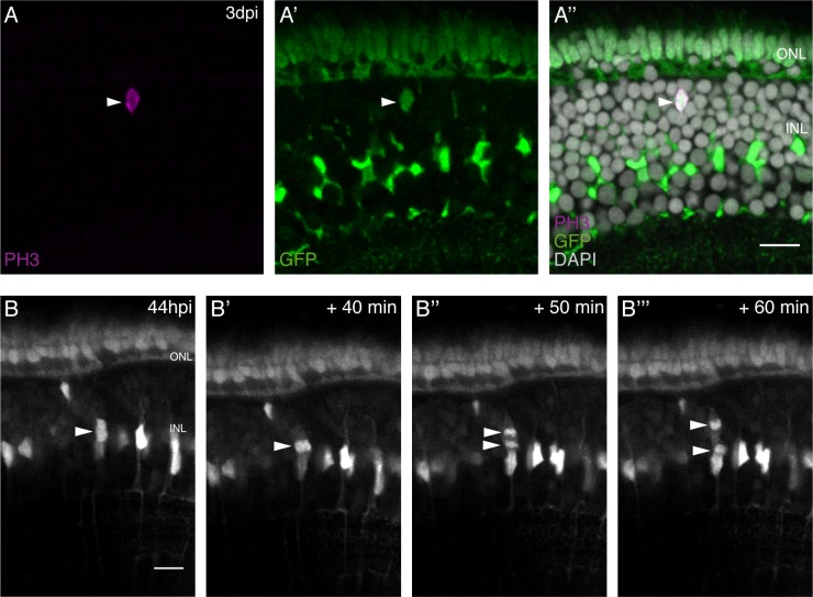
Figure 4.

Figure 4—figure supplement 1. olMG cells divide in various positions in the the INL with an apico-basal spindle orientation.

Figure 4—figure supplement 1.

(A–B) Cryosections of injured hatchling medaka retinae. PH3 stainings (magenta) at 3 days post needle injury show mitotic olMG cells present in different positions in the INL (arrowheads) (n = 4 fish, data obtained from two independent experiments). (C–C’’’) In vivo imaging of hatchling rx2::H2B-eGFP medaka retinae which were injured in the ONL and imaged starting at 44 hpi. An olMG nuclei which starts to condense its chromatin can be detected close to the ONL. The divisions occur in an apico-basal manner (n = 6 fish, data obtained from six independent experiments, 5 out of 6 imaged divisions were apico-basal). Scale bars are 10 μm.
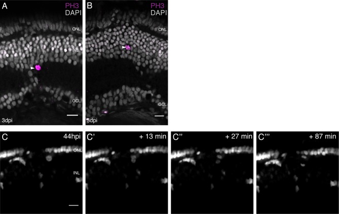
Figure 4—video 1. In vivo imaging of an olMG division after PRC injury.
Download video file (71KB, mp4)
DOI: 10.7554/eLife.32319.017