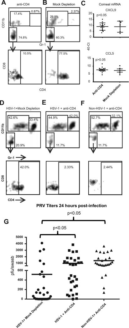
Figure 7. Corneal CD4+ T cells mediate HSV-1-induced resistance to PRV infection.
C57BL/6 mice that received HSV-1 corneal infections 28-34 days previously or non-infected controls received local (subconjunctival) injections of anti-CD4 antibody (CD4-depleted) or control antibody (Mock-depleted). Corneas were excised 3days later and single cell suspensions were stained for CD45, CD4, CD8, CD11b, and Gr-1 and analyzed by flow cytometry (A & B) initially gating on CD45 cells and analyzing for CD11b and Gr-1 (top row) and then gating on the double-negative population and analyzing for CD8 and CD4 (bottom row). The frequency of cells within each area of the flow plot is shown. Alternatively, total RNA was extracted from the HSV-1 infected and CD4-depl or Mock-depleted corneas and CXCL9 and CCL5 chemokine transcripts were quantified by qRT-PCR (C). Data is representative of 1 of 3 experiments in the case of CXCL9 and 1 of 2 experiments regarding CCL5. The HSV-1 infected or mock infected corneas that were CD4-depleted or Mock-depleted were then infected with 1×10⁵ pfu of PRV and 24 hrs later the corneas were excised and single cell suspensions of corneal cells were stained and analyzed by flow cytometry as described above (D-F). Corneal swabs were obtained 24 hrs after PRV infection, infectious PRV was quantified using a viral plaque assay, and results reported as pfu/swab (G). The significance of group differences PRV pfu was assessed by a Kurskal-Wallis test followed by Dunn's multiple comparison test. Data is pooled from three independent experiments.

