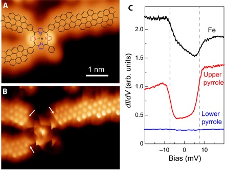
Fig. 2. Imaging and spectroscopy of intact FeTPP connected to cGNRs.

(A) Constant current STM image (with a metal tip) of an FeTPP moiety connected to three cGNRs (Vs = 0.21 V and It = 16 pA). The corresponding structure (as in Fig. 1C) is superimposed. As in the study of Rubio-Verdú et al. (16), the intact FeTPP fragment has a saddle shape, with two lobes due to two pyrrole units pointing upward (red). (B) Constant height dI/dV map of the same structure as in (A) with CO-terminated tip (Vs = 0 mV, Vac = 2 mV rms, and Rt ~ 1 gigaohm). Arrows point to new six-membered rings created after CDH step at the contact region. (C) dI/dV spectra taken on the central Fe atom, on the red pyrrole, and on the blue pyrrole (Rt ~ 50 megaohm on site and Vac = 0.4 mV rms). arb. units, arbitrary units.
