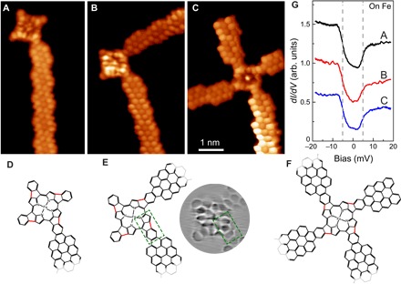
Fig. 3. Imaging and spectroscopy of contacted FeTPP fused with C4 symmetry.

(A to C) Constant height dI/dV maps of planar FeTPP fused to one, two, and four cGNRs, respectively, measured with a CO-functionalized tip (Vs = 0 mV, Vac = 2 mV rms, and Rt ~ 1 gigaohm). All images share the same scale bar. (D to F) Structures corresponding to the hybrids pictured in (A) to (C), respectively. Only a part of the models is shown here for clarity. The red bonds in the structures indicate the clockwise fusion of porphyrin core to the contact phenyl. The green dashed rectangular in(E) highlights the junction structure between FeTPP and cGNRs, with three rings easily recognized in the included Laplacian-filtered image of (B). (G) dI/dV spectra taken on the central Fe atoms of the structures in (A) to (C) (Rt ~ 50 megaohm on site and Vac = 0.4 mV rms). The dI/dV spectra are vertically shifted for clarity.
