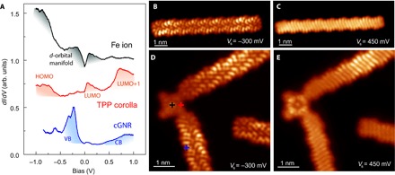
Fig. 5. Comparison of LDOS of pristine and contacted cGNRs.

(A) dI/dV spectra on the center of a contacted porphyrin (top), over a pyrrole group (middle), and over the connected cGNR (bottom). The colored shadows mark relevant bands and resonances, as discussed in the Supplementary Materials and by Rubio-Verdú et al. (16). (B and C) Constant height dI/dV maps of a pristine cGNR measured at the onset values of valence and conduction bands, respectively (Rt ~ 5 gigaohm; the tip was functionalized with CO). (D and E) Constant height dI/dV maps of two cGNRs connected to a planar FeTPP (the structure shown in Fig. 3B) measured as in (A) and (B) at the onset of valence band (VB) and conduction band (CB), respectively. The brighter segment in the upper cGNR branch is caused by the Au(111) herringbone reconstruction underneath.
