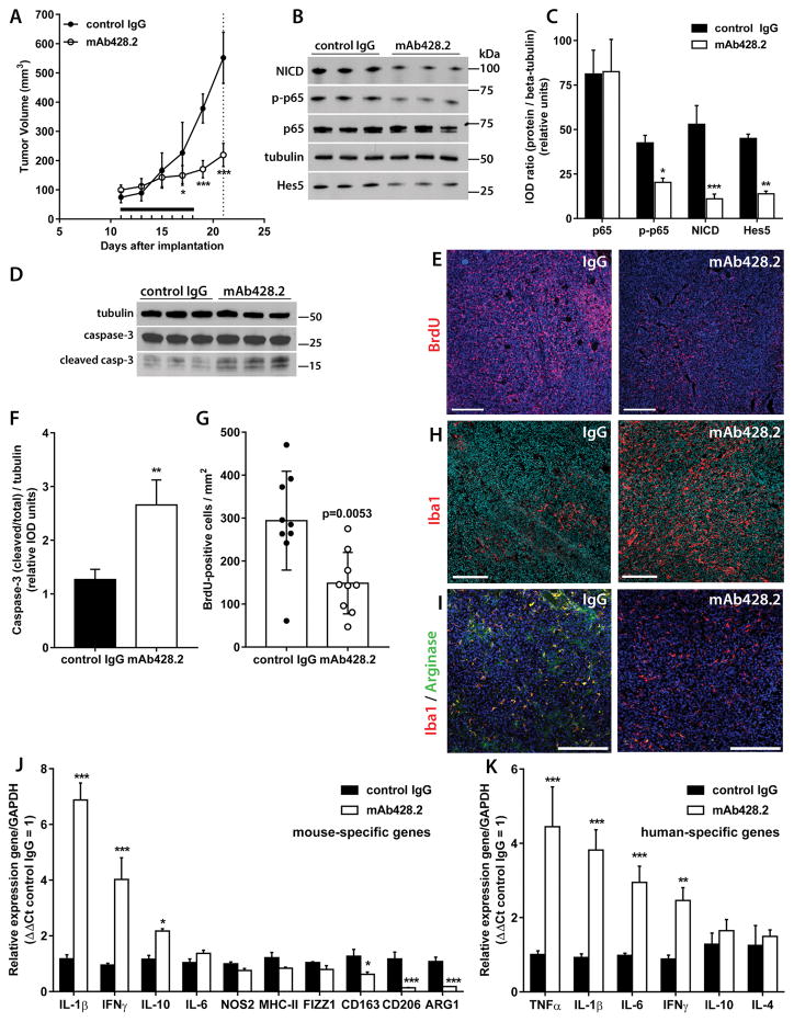
Figure 4. mAb428.2 inhibits tumor growth and increases inflammatory macrophage infiltration in the tumor.
A) Mice carrying bilateral SC GBM34 tumors (N=5/group) were treated with daily injections of mAb428.2 or control IgG for eight days (8 x 30 mg/kg IV, horizontal bar). Tumor growth was significantly inhibited in mAb428.2-treated mice (* p< 0.05; *** p<0.001; repeated measures ANOVA). Animals were euthanized three days after the last injection (dashed line) and their tumors were processed for the rest of the experiments in the figure B–C) Expression of Notch1 Intracellular Domain (NICD) and the transcription factors Hes5 and RelA/p65 (and phospho-p65) was probed by Western blot and quantified by densitometry (IOD: integrated optical density). mAb428.2 treatment significantly reduced expression of NICD, Hes5, and phospho-p65, suggesting inhibition of fibulin-3 effects on Notch and NF-κB signaling (* p<0.05; ** p<0.01; *** p<0.001; Student’s t-test for each marker). D) Expression of full-length and cleaved caspase-3 (casp-3) was probed in three representative tumors per treatment. E) Uptake of BrdU was detected by immunohistochemistry in five tumors per treatment; the images show representative staining results. F) Quantitative analysis of caspase-3 expression from (D) shows increased caspase cleavage in mAb428.2-treated tumors (** p<0.01; Student’s t-test). G) Quantitative analysis of BrdU staining from (E); each dot represents a tissue section. mAb428.2 significantly reduced BrdU uptake (analysis by Mann-Whitney U test). H) Increased macrophage infiltration in mAb428.2-treated tumors, detected by Iba1-positive staining. I) Comparison of tissue sections with similar number of macrophages in both treatments revealed co-expression of Arginase-1 with Iba1-positive cells in control-treated tumors but very low or absent Arginase-1 in macrophages of mAb428.2-treated tumors. J) Tumor treatment with mAb428.2 correlated with increased mRNA expression of the host’s inflammatory cytokines and decreased expression of M2-macrophage markers (CD163, CD206, and ARG1); all genes were detected by qRT-PCR with mouse-specific primers. K) Tumor treatment with mAb428.2 also correlated with increased mRNA expression of inflammatory cytokines in the tumor cells, detected by qRT-PCR with human-specific primers. Results in (J) and (K) were analyzed by Student’s t-test corrected for multiple comparisons (* p<0.05; ** p<0.01; *** p<0.001). Bars in all the histological images: 200 μm.

