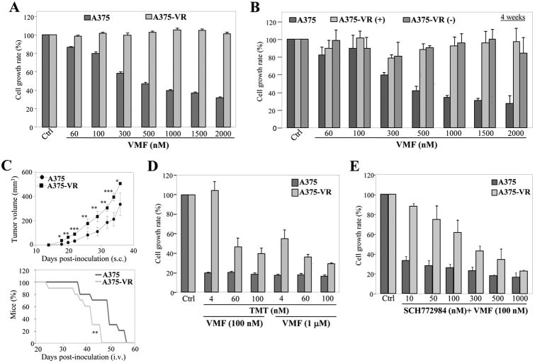
Figure 1. Characterization of vemurafenib-resistant melanoma cells.
(A) Cells were tested in MTT assays (48 h) in the absence (Ctrl) or presence of the indicated concentrations of vemurafenib (VMF) (n=4). (B) A375-VR cells were incubated for 4 weeks without (-) or with (+) VMF (1.3 μM), and subsequently subjected for 48 h to MTT assays as in (A). Parental A375 are shown as control. (C) Cells were subcutaneously (top) or intravenously (bottom) inoculated into NSG mice in the absence of drug treatment, and tumor growth and percentage of alive mice, respectively, were assessed. (n=9-10 mice/condition; ***p<0.001, **p<0.01, *p<0.05). (D, E) Cells were incubated for 48 h without (Ctrl) or with the indicated concentrations of VMF, TMT or SCH772984 (n=3).

